Update on the Morton collection
For updates on the Museum’s work towards the repatriation and reburial of the Morton Collection, please refer to this page.
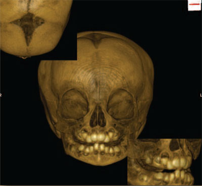
In 2002 a small grant from the University Research Council of the University of Pennsylvania helped launch ORSA—The Open Research Scan Archive—a collection of high-resolution CT scans of human and non-human cranial (cranium and mandible) and post-cranial (everything bone from the neck down) remains. Now supported by a multi-year National Science Foundation grant (number 0447271) to Tom Schoenemann (James Madison University) and Janet Monge (Penn Museum), this online database provides worldwide access to scholars interested in comparative CT scan data.
Since its inception, our CT scanning project has generated over 3,000 scans of the Penn Museum’s skeletal specimens, including two dozen full scans of mummified remains in our Physical Anthropology collections. The project has also scanned numerous CT images of prosimians, monkeys, and apes from the collections of the American Museum of Natural History and Columbia University, and between 2002 and 2007, all 1,200 cranial specimens from the Morton Collection were CT scanned and entered into ORSA. Scholars can now access information about the geographic location from which each cranium was obtained, the identification of the person who collected each specimen and donated them to Morton, and the exact technical specifications of the CT images available.

Yet, besides providing an easily accessible comparative digital archive, what benefits arise from analyzing these CT scans?
One major benefit results from the fact that CT scans give accurate renditions of not only the external surfaces of a bone, but also of the internal bone structure and morphology. For example, recent studies on the origin of bipedalism (walking on two legs) have concentrated on the relationship of this form of walking and the bones found in the region of the middle ear (the malleus, incus, and stapes). These bones, as well as many other structures in the ear, relate to the ability of an individual to maintain their head balance during changes in body position. Since these bones are not visible externally on the skull, scholars must rely on high-resolution CT scans in order to analyze the nature of these bones in different specimens and thereby generate new hypotheses and interpretations about when and how bipedalism developed in our human ancestors.
Another major benefit derived from CT scans is their compatibility with mathematical analysis. Using easily available software, multiple kinds of linear and more complex measurements can be applied to the internal and external features of the CT-scanned bones to generate contours, calculate volumes, and develop even more complex understandings of the geometry of the skull. An example of the latter can be seen in Tom Schoenemann’s work with the Medical Imaging Group that forms part of the Department of Radiology at the Hospital of the University of Pennsylvania. Applying techniques developed in clinical research, they have been able to compare numerous skull specimens to each other, identifying those characteristics that exist as individual elements of human variation from person to person, and then generate a complex 3D model of a ‘generic’ human skull that provides researchers with a baseline from which to compare future specimens. The usefulness of this generic baseline is particularly important when dealing with research questions that focus on how the skulls of males and females differ from each other. Besides providing useful comparative baselines for addressing human evolutionary studies (e.g. are we looking at two different species or are these individuals from one species but representing different sexes), this work also has important applications in forensic anthropology, where the goal is often to factor out individual differences in skulls and to identify broader patterns that might help identify the group to which a specimen belongs (e.g. male or female or this ethnic group or not).
Finally, a third major benefit of CT scans is that they are digital and can be sent electronically to researchers all over the world with the press of a button. This greatly expands the number of researchers in Anthropology, Evolutionary Biology, and the Forensic Sciences with access to the Museum’s Physical Anthropology collections—ORSA is thus a virtual museum of skeletal collections!
